Scientists at Oak Ridge National Laboratory (ORNL) have pioneered an advanced method to observe the motion of domain walls in ferroelectric materials with exceptional precision.

Understanding Ferroelectricity
Ferroelectricity is a characteristic found in certain insulating materials or dielectrics, where there is a natural separation of positive and negative charges. This separation leads to spontaneous electric polarisation, creating distinct positive and negative regions within a crystal.
This polarisation can be reversed by applying an external electric field. The term “ferroelectric” is inspired by “ferromagnetism”—a phenomenon where magnetic domains spontaneously align. In a similar fashion, electric dipoles in ferroelectric materials naturally align within domains.
Common ferroelectric substances include barium titanate (BaTiO₃) and Rochelle salt. These materials form domains with uniformly aligned dipoles, and their orientation can be altered with strong electric fields.
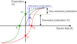
The lag in the dipoles’ reorientation during polarity reversal is known as ferroelectric hysteresis, analogous to the hysteresis observed in magnetic materials.
Ferroelectric properties diminish beyond a specific temperature known as the Curie temperature, where thermal energy disrupts the orderly dipole alignment.
Domain Walls in Ferroelectric Materials
Domain walls are interfaces between regions with different polarisation directions within a ferroelectric crystal. These boundaries often exhibit distinct electrical or magnetic behaviours compared to the adjacent domains.
For example, some domain walls may become electrically conductive even if the surrounding material remains insulating, or display magnetic properties in otherwise nonmagnetic regions.
Because of their unique behaviour, domain walls are promising components for future nanoelectronics, potentially enabling low-power memory storage, sensors, and signal processing technologies.
ORNL’s Innovative Imaging Method
The newly developed technique—Scanning Oscillator Piezoresponse Force Microscopy (SO-PFM)—allows scientists to capture both gradual and sudden movements of domain walls under swiftly changing electric fields.
Unlike previous methods that provided only static images before and after changes occurred, this technique offers real-time, dynamic imaging. This advancement allows researchers to visualize how domain walls shift over time and determine the energy required to move them.
The process integrates precisely timed electronic controls with atomic force microscopy (AFM), unlocking new possibilities for observing live domain wall dynamics that were previously inaccessible.


